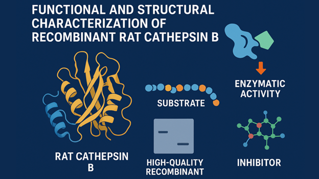
Cathepsin B is a cysteine protease that belongs to the cathepsin family of lysosomal proteases. Primarily found within lysosomes and endosomes, Cathepsin B performs proteolytic activities, including the degradation of intracellular proteins and both exopeptidase and endopeptidase activities.
This dual enzymatic capability is critical for diverse biological processes such as:
- Routine cellular cleanup
- Degradation of complex host blood proteins
Recombinant Production, Processing, and Characterization
Obtaining reliable structural data always requires high-quality recombinant rat Cathepsin B. This is why the production must address all the post-translational modifications and activation to ensure that the final product mimics the native enzyme.
Expression Systems and Manufacturing Standards
Recombinant rat Cathepsin B is typically expressed in heterologous systems with mammalian cell lines. These cells facilitate the proper folding and incorporation of mammalian-specific post-translational modifications (PTMs). The result is a protein with characteristics more closely resembling those of the native enzyme.
It is synthesized as a preproenzyme. The specific recombinant protein construct often corresponds to the mature protein sequence. Engineering it with an affinity tag streamlines purification processes.
Post-Translational Processing and the Role of Glycosylation
The native procathepsin B zymogen undergoes co-translational translocation into the rough endoplasmic reticulum. This is followed by N-glycosylation at potential sites within the propeptide and the heavy chain.
Studies using yeast-expressed enzymes demonstrate that purified fractions of Cathepsin B exhibiting the highest levels of glycosylation display reduced activity. This confirms that glycosylation may influence stability and protein interactions.
However, the polypeptide structure itself dictates the core catalytic function. Regardless of the minor variations in N-glycan patterns from different expression systems, the characterization of recombinant rat cathepsin B serves as a true representative model for detailed kinetic and structural studies.
Quality Metrics and Activation Protocol
The reliability of the enzyme as a research reagent depends on quality control. High-grade RRCathB can have a purity exceeding 95% and low endotoxin levels (<1.0 EU/μg). Fluorogenic peptide substrates enable the quantification of biological activity.
High-quality RRCathB is expected to show activity greater than 2,500 pmol/min/μg against Z-LR-AMC. Since it is produced as a proenzyme, it must first be activated in an acidic, reducing buffer. This step allows accurate kinetic measurements and replicates the enzyme’s natural lysosomal processing.
High-Resolution Structural Architecture
The Mechanism of the Occluding Loop
Cathepsin B gets its structural element and unique dual activity from the ‘occluding loop,’ an insertion loop consisting of approximately 18 amino acids. The loop physically blocks a portion of the substrate-binding cleft, forcing the enzyme toward peptidyldipeptidase (exopeptidase) activity.
pH dynamically regulates the conformational stability of the occluding loop. At acidic pH, the formation of two key salt bridges (His110–Asp22 and Arg116–Asp224) stabilizes the conformation. This locks the occluding loop, restricting the enzyme’s activity by preventing full access to the enzyme’s binding pocket.
At acidic pH, stabilization of the occluding loop in its closed conformation restricts substrate access and predominantly drives exopeptidase activity, resulting in the removal of two residues from the C-terminus.
The displacement (mobility) of the occluding loop in a neutral pH environment is the structural change that permits greater access to the full substrate-binding cleft, thereby increasing the potential for endopeptidase activity while its major function remains exopeptidase.
Substrate Recognition Subsites
The substrate recognition subsites (S2–S1–S1′–S2′) determine the substrate specificity of Cathepsin B. These binding sites attach to amino acids near the cleavage point to control catalytic efficiency and selectivity.
S1 Subsite prefers hydrophobic or basic amino acids such as leucine or arginine.
S2 Subsite is a conserved pocket located near the catalytic residue Cys29. It is responsible for recognizing the cleavage site.
S1′ and S2′ Subsites are flexible regions allowing the enzyme to accommodate a wide range of substrate sequences.
Applications of Recombinant Rat Cathepsin B in Drug Discovery
Screening and Structure-Based Design
High-throughput screening uses the characterization of recombinant rat cathepsin B to evaluate large compound libraries for potential inhibitory activity.
Crystallographic data from the RRCathB complexed with inhibitors is critical in structure-based drug design (SBDD). Analysis confirms that inhibitor binding induces very little conformational change in the core structure of the enzyme. This structural stability simplifies the drug design process.
Role in Targeted Drug Delivery
Targeted drug delivery utilizes Cathepsin B as a key molecular target. Its overexpression in breast, liver, and colorectal cancers provides a mechanism for drug activation through tumor-specific triggers.
Inhibitor Selectivity and Translational Challenges
Changes in the structure of Cathepsin B depend on pH. It is also similar to other cysteine cathepsins. This makes it challenging to develop selective inhibitors. The occluding loop provides some selectivity, but inhibitors are often less effective in living systems.
Find a Home-Based Business to Start-Up >>> Hundreds of Business Listings.


